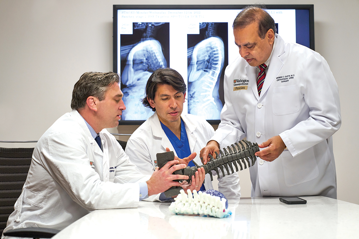
MUNISH GUPTA, MD, USES A 3D MODEL OF A PATIENT’S SPINE AS AN AID IN PLANNING SURGERY.
Photography by Jennifer Sliverberg
BY STEPHANIE STEMMLER
At WashU Medicine’s Living Well Center, people scheduled for surgery to correct spinal problems gather to learn how they can play an important role in their own recovery. In this special program designed by WashU Medicine orthopedic spine surgeons and neurosurgeons, attendees work with a nurse practitioner, a nutritionist and therapists in a newly launched Spine Surgery Optimization Program that includes health topics such as smoking cessation, good nutrition, coping mechanisms for pain and other topics.
“We are helping these patients prepare for a life-changing surgery,” says Brian Neuman, MD, WashU Medicine orthopedic spine surgeon at Barnes-Jewish Hospital and chief of the Section of Spine Surgery in the Department of Orthopedics. “We begin as far out as two months prior to surgery because we want people to be in the best physical and mental health possible before their procedures.”
Up to 50% of people with spinal deformity can have surgical or medical complications following surgical treatment, notes Neuman, which can affect a patient’s overall health and mobility. However, research has shown that if patients are healthier and more mobile prior to surgery, they have better outcomes and fewer complications in their recovery over the long term.
WashU Medicine neurosurgeon and researcher Jacob Greenberg, MD, MSCI—working with WashU Medicine orthopedic spine surgeon Munish Gupta, MD, MBA, and Neuman—led a pilot study with a small group of people with spinal deformity to determine whether a patient’s mobility before surgery affected outcomes after surgery. The results of this study resulted in the formation of the Spine Surgery Optimization Program. Now, a two-year study co- led by WashU Medicine orthopedic spine surgeons and neurosurgeons is under way to evaluate the program’s effectiveness.
“We’re looking at a variety of factors that can affect outcomes, including social support, medical concerns, co-morbidities, bone structure, lifestyle issues and fortitude,” explains Neuman. “We have known for years that these things can impact surgical outcomes. Now we’re pushing hard to make use of that information so we can obtain the best possible outcome for the people we treat.”
Spinal deformity: an anatomy lesson
The human spine includes 33 vertebrae, which run from the bottom of the skull to the pelvis. In most people, the spine curves in four places: the neck (cervical), back (thoracic), lumbar (lower back) and sacrum, located just above the pelvis.
Some conditions cause the spine to bend in other places; for instance, from side to side, front to back, or in a twisted combination of the two.
Common spinal problems that contribute to different curvatures include:
- - Scoliosis: a lateral or side-to-side curve or rotation of the spine that can give the spine an S-shaped or C-shaped appearance.
- - Kyphosis: an excessive forward curve of the spine along the upper back, which causes a person to appear hunched over.
- - Lordosis: an inward curve of the spine along the lower back, sometimes known as swayback.
- - Spondylolisthesis: a forward slipping of the vertebral bone in the spine
Fractures and spinal tumors also can cause problems with curvature. Regardless of the cause, such deviations in the spine can result in painful problems that can progressively worsen. Many people with these conditions experience difficulty walking and have nerve damage.
Over the past few decades, treatments to fix these kinds of spinal differences have included the use of steel wires, as well as screws, plates and steel rods. Surgery using these technologies was designed to correct the abnormal curve and stabilize the spine from top to bottom. But these procedures couldn’t replicate the natural, undulating curve of a normal spine. Inserting a rod into the back could straighten a curve, but after surgery the spine might have no curve at all. This limitation often caused people to require a second, or revision, surgery.
Advances bring promise
Classes like those that are part of the Spine Surgery Optimization Program have improved outcomes for complex spinal surgery. Additionally, new techniques and technologies are advancing such treatment, offering better quality of life after surgery.
“We have known for many years that we can remove a wedge-like portion of bone from the back of the lumbar spine to correct flatback syndrome, but rather than use a single rod, we now use multiple, shorter rods that allow for improved outcomes and decreased failure rates,” Gupta says. That innovation, coupled with advances in computer imaging and 3D modeling—plus real-time tracking of the alignment changes made during surgery—have improved outcomes.
Additional advances include the use of recombinant spinal fusion materials, including bone morphogenetic protein, or BMP. These materials are genetically engineered and, when used as a substitute for bone grafts in spinal surgery, can offer higher success rates in fusing to existing bone.
The techniques used during spine surgery also have evolved over the years. Traditionally, surgeons accessed the spine through the back. Today, advances allow surgeons to access the spine from the front or side, depending on the type and severity of curvature. As a result, Gupta notes, surgeons can correct more severe degrees of curvature.
Precise and personalized treatment
Perhaps the most fascinating changes in spinal surgery are possible thanks to sophisticated computer software technology and high-resolution imaging. Surgeons now routinely take multiple images or scans of a patient’s spinal anatomy and integrate them into detailed 3D models that can be manipulated on screen and studied in all directions for greater precision in pre-surgical planning.
“And that same technology can guide us in making customized rods that match in size and curvature a patient’s unique anatomy,” says Neuman. “We really have achieved precision medicine for spinal corrections.”
New technology also allows surgeons to use full-length digital X-rays of the spine, which are taken throughout surgery to ensure the best spinal correction is achieved. At Barnes-Jewish Hospital, WashU Medicine surgeons wear augmented-reality glasses that allow layering of enhanced spinal images over real-time surgical views. This enhanced view provides greater accuracy in placing grafts and correcting curvature. Technology also gives the surgeon neuro-monitoring capability, which ensures that nerves within the spinal column are not adversely impacted during surgery.
Collaboration is key
People needing surgery for complex spine problems also benefit from collaboration between orthopedic and neurosurgery specialists.

IMAGE 1: X-RAY SHOWING A CHONDROSARCOMA TUMOR OF THE SPINE; IMAGE 2: 3D RENDERING OF AN IMPLANT DESIGNED TO FIT THE SPINE AFTER REMOVAL OF TUMOR; IMAGE 3: X-RAY SHOWING FINAL PLACEMENT OF THE IMPLANT.
Images courtesy of WashU Medicine
“The vast majority of spine pathologies could be managed either by neurosurgeons or by orthopedic spine surgeons,” says Camilo Molina, MD, FAANS, WashU Medicine neurosurgeon and spine specialist at Barnes-Jewish Hospital and director of the Neurosurgery Spinal Deformity and Spinal Oncology Service. “But to best treat people with unique, complex problems, we work side by side.” Molina notes that, thanks to consultation meetings attended by both specialties, preoperative planning is more detailed and robust. “We can be more efficient in the operating room as a result, and we are able to use advanced treatment options because of this collaboration.”
Working together, Molina and colleague Matthew Goodwin, MD, PhD, WashU Medicine orthopedic spine surgeon at Barnes-Jewish Hospital, are forging a new frontier in surgery to treat spinal tumors. Removal of these types of tumors is highly technical; to achieve the best outcome, the tumor needs to be removed in its entirety, in one piece. To achieve this goal, Molina and Goodwin use 3D modeling before surgery to plan every step. The technology has enabled them to expand what they can remove in terms of the location of the tumor and its size. Both Molina and Goodwin treat patients with cancerous spinal tumors at Siteman Cancer Center, based at Barnes-Jewish Hospital and WashU Medicine.
This type of surgery presents a second challenge: After removal of the tumor, the spine may need to be reconstructed. Cancer can eliminate bone, leaving empty spaces in the skeleton where tumors used to be. To help solve this problem, Molina is evaluating the use of 3D-printed, patient-specific implants to reconstruct spine and bone after tumor removal. The process is complex.

A MULTIDISCIPLINARY TEAM COLLABORATES ON PLANNING COMPLEX SPINAL SURGERY
“We pre-design an implant based upon the planned removal of a spinal tumor,” explains Molina. “After the tumor is removed, we use CT scans and 3D modeling to identify where bone is lost and needs to be replaced. We then put in temporary fixation devices. After the first surgery, we manufacture a titanium implant, sterilize it and place it in the spine in the second phase of this two-part procedure.”
To date, Molina and Goodwin have performed six of these procedures as part of an ongoing study. Molina notes that more research is needed; currently, this innovative process is approved by the Food and Drug Administration (FDA) only on a case-by-case basis. Molina notes that new technology and new techniques have significantly improved treatment for complex spinal problems, calling advances in both pre- and post-surgery care “game-changers.” It’s also true, he adds, “that collaboration between neurosurgery and orthopedics benefits patients. When we work together, we can push the field farther to make surgery for complex deformities better.”
Learn more about spinal surgery.