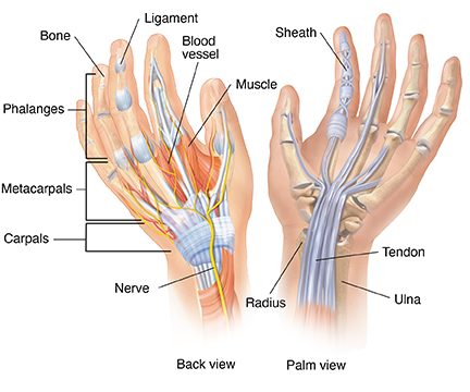
Anatomy of the Hand
The hand is composed of many different bones, muscles, and ligaments that allow for a large amount of movement and dexterity.

There are three major types of bones in the hand itself:
-
Phalanges. These 14 bones are found in the fingers of each hand and also in the toes of each foot. Each finger has three phalanges (the distal, middle, and proximal). The thumb only has two.
-
Metacarpal bones. These five bones make up the middle part of the hand.
-
Carpal bones. These eight bones create the wrist. The two rows of carpal bones are connected to two bones of the arm—the ulna bone and the radius bone.
Numerous muscles, ligaments, tendons, and sheaths can be found within the hand. The muscles are the structures that can contract, allowing movement of the bones in the hand. The ligaments are fibrous tissues that help bind together the joints in the hand. The sheaths are tubular structures that surround part of the fingers. The tendons connect muscles in the arm or hand to the bone to allow movement and typically pass through the sheaths.
In addition, there are arteries, veins, and nerves within the hand that provide blood flow and sensation to the hand and fingers.